Knee Arthroscopy (Key Hole Surgery)
What is Arthroscopy?
Arthroscopy is keyhole surgery used to diagnose and treat problems within the knee joint.
Arthroscopy equipment is small, so only small cuts in the skin are needed. This has the advantage of less pain after the operation, faster healing time, and lower risk of infection.
Causes
The procedure is used to treat a number of conditions, including tears of the cartilage, (meniscus), removing fragments of loose bone or cartilage, drilling of bone to promote healing of cartilage and treatment of an anterior cruciate ligament (ACL) tear.
The most common reason for knee arthroscopy surgery done by Mr Mahaluxmivala is a torn meniscus (shock absorbing cartilage) of the knee. Both the cartilage on the inside and outside of the knee can be torn and both can be suitable for knee arthroscopy.
Mr Mahaluxmivala explains however that this is one of the operations where “patient selection” is vital for this surgery to have a successful outcome. Insurance companies have strict criteria for allowing this surgery to be performed and this is done to make sure unnecessary surgeries do not occur.
Symptoms
- The best results of this surgery occur when the patient gives a clear history of twisting or injuring his or her knee, which brought on the symptoms rather than a gradual onset.
- The patient’s main complaints will be locking and giving way (mechanical symptoms) rather than unrelenting pain. The patient may be a normally active sporty person with no x-ray evidence of arthritis setting in the knee.
- The patient may have pain all over the knee or deep within the knee.
- More commonly, the patient will complain of pain on the inner aspect or outer aspect of the knee.
- The pain and acute catching or locking in the knee will be worse when the patient walks on an irregular surface. It is also worse when he or she pivots or twists on the knee. A common complaint is, if the patient hits the foot accidentally against any object, there will be a sharp pain referred to the inner aspect of the knee area
- The knee may feel swollen and inflamed initially when symptoms commence.
Diagnosis
- Diagnosis is made from the history and complete examination.
- An x-ray of the knee which includes three specialised views including standing views is needed.
- A MRI scan is done which will prove a tear of the cartilage or other ligament problems. It will also pick up arthritis in the knee which has not shown up on x-rays.
Mr Mahaluxmivala will show the patient the images of his or her x-rays and MRI on the monitor in the consulting room demonstrating the problem which necessitates the need for a knee arthroscopy.
When Is Arthroscopy Or Key Hole Surgery Done?
Treatment
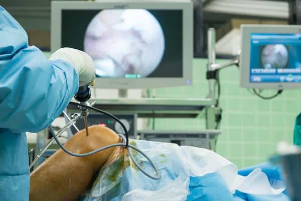
During the surgery, a small cut, a few millimetres long, is made in the skin next to the joint so that an arthroscope (a metal tube attached to a camera and a light source) can be inserted.
One or more additional incisions will also be made and the integrity of the anterior cruciate ligament can be confirmed and any irregular areas of cartilage can be smoothened or removed with high-performance mini shavers and burrs. Drilling of the bone is performed if needed.
The arthroscope sends images to a video screen monitor. These images will be saved and shown to the patient.
The time for this type of knee arthroscopy surgery is less than an hour, but Mr Mahaluxmivala reassures relatives not to expect the patient back in the ward in this short time span. The reason is that a lot of care and time is taken before surgery in preparing the patient for anaesthesia, along with safe and correct positioning of the patient.
This along with the recovery time where the patient is monitored very closely after the operation means it will be longer before the patient returns to the ward.
Our Clinic is based at the Rivers Private Hospital in Sawbridgeworth, Hertfordshire.